Defibrillator (ICD) Placement
A defibrillator is an implantable medical device that (1) acts as a pacemaker capable of delivering electricity to the heart to trigger it to beat, and (2) if necessary, shocks the heart out of life-threatening heart rhythms. It is this second feature, or the ability to shock the heart, that predominantly differentiates a defibrillator from a standard pacemaker.
Certain individuals (e.g. those with severe congestive heart failure) are prone to have life-threatening heart rhythms and may be candidates for placement of a defibrillator, also known as an automatic implantable cardioverter defibrillator (ICD). All defibrillators, as explained above, serve a dual role, capable of being both a pacemaker and a defibrillator (shocking) device when needed.
Understanding the Defibrillator Placement Procedure
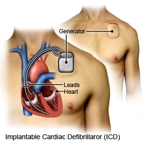
 Following patient sedation, a small incision (approximately 2-3 inches) is made beneath the collar bone. Electrodes (termed “leads”) are then inserted into the subclavian vein and passed inside this vein to the heart (see image on the right). Electrodes have small screw-like coils on their tips that enable them to be secured into place within the heart muscle. The electrodes are then attached to the defibrillator generator which is placed underneath the skin. The skin surface is closed with sutures.
Following implantation, defibrillator function is closely monitored on follow-up visits. During device checks, which may be performed in the physician’s office or even at home with telephonic monitoring, detailed information about both the defibrillator (e.g. battery life or frequency of pacing) and the intrinsic heart (e.g. underlying rhythm) is able to be obtained and helps with ongoing management.
Blending cutting-edge technology with traditional compassion.
Call for an Appointment
941-421-0580
